This is the second entry continuing with the summary of the main research findings on gender differences in the functioning and structure of the brain, including biochemical processes. This entry will concentrate on the differences between the hemispheres and connective tissue of the brains of males and females, as well as their subcortical brain regions (the hypothalamus will be addressed in the next entry).
Size Difference in the Hemispheres and Connective Tissue
The neocortex is made up of two halves called hemispheres. Hemispheres have conlateral control over the body, meaning that each hemisphere controls the side of the body opposite to it.
Hemispheres
Nearly all studies show that females have more evenly proportional hemispheres, while males have a greater disparity in the size of their hemispheres, with the right hemisphere being larger. When right hemispheres are compared, studies suggest that males usually have larger ones than females.
Size of the Corpus Callosum

The corpus callosum is the tissue that connects the two hemispheres of the brain as well as connecting the neocortex to the subcortical region of the brain.
Considerable research has shown that the corpus callosum is usually larger in females than it is in males, specifically the midsagittal corpus callosum cross-sectional area.
Interestingly, it has also been found to be larger in musicians, in left-handed and ambidextrous people, while dyslexic children tend to have smaller and less developed corpus callosums.
Size of Broca’s Area

There is a region on the left frontal lobe, called Broca’s area, that is instrumental in the production of speech in human beings.
Two studies conclude that Broca’s area is significantly larger in females than it is in males.
Size of Wernicke’s Area
Located in the posterior portion of the neocortex’s left hemisphere, Wernicke’s area serves to interpret linguistic communication (written and spoken language).
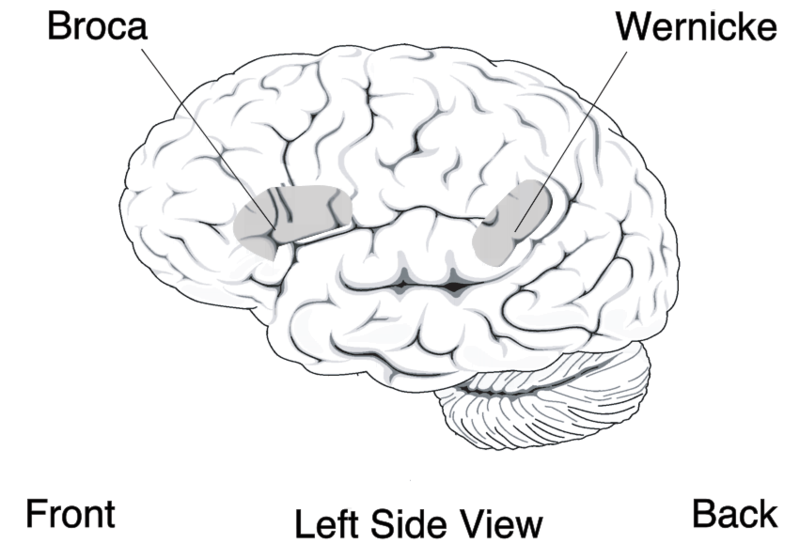
One study shows that Wernicke’s area is on average larger in females.
Subcortical Brain Regions
All the parts of the brain apart from the neocortex are called the subcortex.
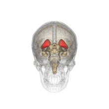
Size of the Amygdala
The amygdala is part of the limbic system, or the emotion-regulating part of the subcortex in mammals. There are two of these almond-shaped clusters of nuclei, each in one of the temporal lobes. The amygdala play a role in the processing of memory, decision-making, and emotional responses.
The limbic system includes a variety of functions, including emotion, behaviour, motivation, long-term memory, and the sense of smell.
In humans, while some studies have shown no significant difference in the size of the amygdala between males and females, the majority have concluded that all or parts of the amygdala are larger in males than in females, from ages 7 to 11 (reference), as well as in adulthood (reference).
When men and women were shown horror movies (emotionally provocative in the negative sense) and neutral movies
Consistent with suggestions from several previously published studies, enhanced activity of the right, but not the left, amygdala in men was related to enhanced memory for the emotional films. Conversely, enhanced activity of the left, but not the right, amygdala in women was related to enhanced memory for the emotional films. These results demonstrate a clear gender-related lateralization of amygdala involvement in emotionally influenced memory, and indicate that theories of the neurobiology of emotionally influenced memory must begin to account for the influence of gender. (reference)
The right amygdala is linked with taking action and negative emotions, which may explain why males tend to respond to emotionally stressful stimuli physically.
The left amygdala allows for the recall of details, but it also results in more thought rather than action in response to emotionally stressful stimuli, which may explain the absence of physical response in women.
According to responses on Izard scales, electrical stimulations of the right amygdala induced negative emotions, especially fear and sadness. In contrast, stimulations of the left amygdala were able to induce either pleasant (happiness) or unpleasant (fear, anxiety, sadness) emotions. Unpleasant states induced by electrical stimulations were accompanied by an increase in EMGc activity. In addition, when emotional changes were reported after electrical stimulation, SCR amplitude for the positively valenced emotions was larger than for the negative ones. These findings provide direct in vivo evidence that the human amygdala is involved in emotional experiences and strengthen the hypothesis of a functional asymmetry of the amygdala for valence and arousal processing.
(reference)
Another study explains that:
Growing evidence from recent neuroimaging studies points to a new, expanded role for the amygdala as a critical structure that mediates sex differences in emotional memory and sexual responses. This review highlights current findings from studies of sex differences in human amygdala response during emotion-related activities, such as formation of emotional memories and sexual behavior, and considers how these findings contribute to the understanding of behavioral differences between men and women. Clinical implications for the understanding of sex differences in the prevalence of affective and anxiety disorders are discussed, and future directions in the study of the amygdala’s role in human sex differences are outlined. (reference)
and concludes that, as a result, women on average tend to retain stronger memories for emotional events than men.
Some recent studies suggest possible correlations between brain structure, including differences in hemispheric ratios and connection patterns in the amygdala, and sexual orientation. Homosexual men tend to exhibit more feminine patterns in the amygdala than heterosexual males do, just as homosexual females tend to show more masculine patterns in the amygdala than heterosexual women do. It was observed that amygdala connections were more widespread from the left amygdala in homosexual males, as is also found in heterosexual females. Amygdala connections were more widespread from the right amygdala in homosexual females, as in heterosexual males.
Here are a couple of abstracts from such studies
PET and MRI show differences in cerebral asymmetry and functional connectivity between homo- and heterosexual subjects:
Cerebral responses to putative pheromones and objects of sexual attraction were recently found to differ between homo- and heterosexual subjects. Although this observation may merely mirror perceptional differences, it raises the intriguing question as to whether certain sexually dimorphic features in the brain may differ between individuals of the same sex but different sexual orientation. We addressed this issue by studying hemispheric asymmetry and functional connectivity, two parameters that in previous publications have shown specific sex differences. Ninety subjects [25 heterosexual men (HeM) and women (HeW), and 20 homosexual men (HoM) and women (HoW)] were investigated with magnetic resonance volumetry of cerebral and cerebellar hemispheres. Fifty of them also participated in PET measurements of cerebral blood flow, used for analyses of functional connections from the right and left amygdalae. HeM and HoW showed a rightward cerebral asymmetry, whereas volumes of the cerebral hemispheres were symmetrical in HoM and HeW. No cerebellar asymmetries were found. Homosexual subjects also showed sex-atypical amygdala connections. In HoM, as in HeW, the connections were more widespread from the left amygdala; in HoW and HeM, on the other hand, from the right amygdala. Furthermore, in HoM and HeW the connections were primarily displayed with the contralateral amygdala and the anterior cingulate, in HeM and HoW with the caudate, putamen, and the prefrontal cortex. The present study shows sex-atypical cerebral asymmetry and functional connections in homosexual subjects. The results cannot be primarily ascribed to learned effects, and they suggest a linkage to neurobiological entities. (reference)
Sexual Differentiation of the Brain and Behavior:
During the intrauterine period the human brain develops in the male direction via direct action of a boy’s testosterone, and in the female direction through the absence of this hormone in a girl. During this time, gender identity (the feeling of being a man or a woman), sexual orientation, and other behaviors are programmed. As sexual differentiation of the genitals takes places in the first 2 months of pregnancy, and sexual differentiation of the brain starts during the second half of pregnancy, these two processes may be influenced independently of each other, resulting in transsexuality. This also means that in the case of an ambiguous gender at birth, the degree of masculinization of the genitals may not reflect the same degree of masculinization of the brain. Differences in brain structures and brain functions have been found that are related to sexual orientation and gender. (reference)
Sexual Orientation and its Basis in Brain Structure and Function:
Current evidence indicates that sexual differentiation of the human brain occurs during fetal and neonatal development and programs our gender identity—our feeling of being male or female and our sexual orientation as hetero-, homo-, or bisexual. This sexual differentiation process is accompanied by many structural and functional brain differences among these groups (1). In previous studies (2, 3), the Savic laboratory detected a sex-differentiated activation of the anterior hypothalamus in heterosexual men (HeM) and heterosexual women (HeW) and a sex-atypical, almost reversed, pattern of activation in homosexual men (HoM) and homosexual women (HoW). The hypothalamus (Fig. 1) is a small brain area located under the anterior commissure that is involved in many different functions, including reproduction. These observations raised several questions, one of which was whether the sexual dimorphisms described could be sex-atypical in homosexual subjects even with respect to factors not directly associated with reproduction. In a recent issue of PNAS, Savic and Lindström (4) reported that hemispheric ratios, as well as patterns of amygdala connectivity, were sex-atypical in homosexual individuals, with HoM exhibiting more female patterns than HeM and HoW showing more male-like features than HeW. Whether the observed sex-atypical characteristics are the result of processes that occur during the fetal or neonatal periods, as is the case with gender identity and sexual orientation, is an open question. The excellent imaging research of Ivanka Savic’s group in past years has provided strong evidence for structural and functional brain differences related to gender and sexual orientation. […](reference)
Size of the Basal Ganglia
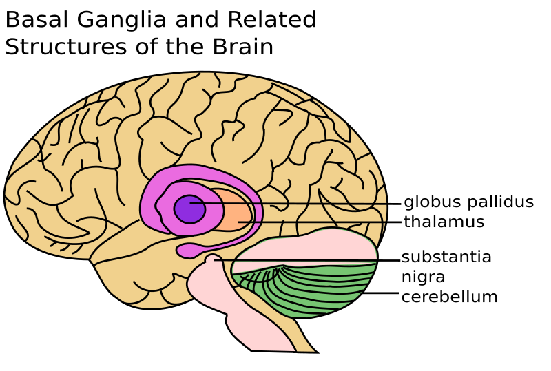
The basal ganglia are a cluster of nerve cells at the base of the brain, making up the putamen, caudate, globes pallidus, and substantia nigra. Their main function is related to the regulation of motor function, procedural learning, habit learning, eye movement, cognition and emotion. Dysfunction of the basal ganglia results in a wide range of neurological and behavioural conditions, including Tourette syndrome, obsessive-compulsive disorder, addiction, Parkinson’s, Huntington’s disease, dystonia and hemiballismus.
One study concludes that the basal ganglia is larger in males than in females.
Size of the Bed Nucleus of the Stria Terminalis
The bed nucleus (of the stria) terminalis is a small portion of the hypothalamus connected to the amygdala that is found in some mammals, including humans. Its activity correlates with anxiety in response to threats, and it seems to regulate the response to acute stress of more than 10 minutes.

All research has shown that this portion of the hypothalamus is larger in males than in females. On average, it is twice as large in men as in women, and contains twice the number of somatostatin (regulating growth hormone…) neurons, regardless of sexual orientation.
The stria terminalis is sexually dimorphic, which means that it is characteristically different in the two sexes of a species. Studies have shown that while this area of the brain is essential for sexual behaviour, its size was not influenced by sex hormones in adulthood and was independent of sexual orientation, and that “in transsexuals, sexual differentiation of the brain may go into opposite directions and point to a neurobiological basis of gender identity disorder”. In other words, gender identity would be determined before birth… See this study and this study.
The larger bed nucleus of the stria terminalis found in men (including transgender men) reduces the startle response in men and may be responsible for the higher incidence of specific phobias in women, usually documented in psychiatry.
Sexual maturity and development may be impacted by deficits in the bed nucleus of the stria terminalis during childhood. For instance, a study of the neurobiological factors of pedophilic subjects showed
a significant decrease of right amygdalar volume, compared with healthy controls. We observed reduced gray matter in the right amygdala, hypothalamus (bilaterally), septal regions, substantial innominata, and bed nucleus of the striae terminalis. In 8 of the 15 perpetrators, enlargement of the anterior temporal horn of the right lateral ventricle that adjoins the amygdala could be recognized by routine qualitative clinical assessment. Smaller right amygdalar volumes were correlated with the propensity to commit uniform pedophilic sexual offenses exclusively. (reference here)
Allow me to quote the conclusions of this same article:
Conclusions Pedophilic perpetrators show structural impairments of brain regions critical for sexual development. These impairments are not related to age, and their extent predicts how focused the scope of sexual offenses is on uniform pedophilic activity. Subtle defects of the right amygdala and closely related structures might be implicated in the pathogenesis of pedophilia and might possibly reflect developmental disturbances or environmental insults at critical periods.
The sexual abuse of children is a major public health and criminological issue. Although, at present, no reliable data are available on the prevalence of pedophilia, it was recently reported that about 1 in 12 children in the United States has been sexually approached by an adult.1 In contrast to tolerance of such behavior in ancient Greece, these days pedophilia is not socially acceptable. Instead, it is considered a paraphilia and has been operationally defined as a psychiatric disease.2 The diagnostic criteria require that an adult experiences a sustained sexual attraction to prepubescent children and has acted on it or feels distressed by this sexual urge. Many attempts have been made to identify a pattern of neurocognitive and neurobiological markers reliably related to pedophilia. A recent meta-analysis by Cantor et al3 revealed overall impaired intelligence in sexual offenders of children. However, no pattern of neurobiological characteristics has been documented consistently in a large sample of such offenders.1 A variety of risk factors have been linked to increased incidence of pedophilic behavior: environmental risk factors (eg, childhood sexual abuse4 and an inadequate attachment style resulting from a dysfunctional family5), neurobiological factors (eg, hormonal alterations1,6), and neurodevelopmental disturbances,7 as well as acquired organic conditions, mainly involving frontal and temporal regions.8,9
Previous imaging studies comparing groups of pedophilic patients with control groups are scarce. Functional neuroimaging studies using positron emission tomography have indicated a role for frontal and temporal abnormalities in pedophilia.9,10 Structural studies based on computed tomography scans and their qualitative assessment have provided inconsistent results. Some have suggested abnormalities of temporal11,12 as well as frontal12 regions. Others have failed to find a significant association with structural brain anomaly.13
Whereas little is known about brain function in pedophilia, the structures involved in sexual behavior in other populations have been extensively studied. Frequently, patients with lesions of the anterior temporal lobes and the frontal lobes show changes in their sexual behavior.8,14 These can vary from slight changes in sexual drive to full-blown symptoms of the Klüver-Bucy syndrome, which consist of hyperorality, so-called psychic blindness, hypersexuality, and changes in sexual preference.15 Furthermore, it has been established in animals that a densely interconnected neural network involving the amygdala, hypothalamus, septal area, and cell groups in the adjacent substantia innominata plays a key role in determining sexual and mating behavior.16,17 Functional imaging studies in healthy humans show that the homologous human structures are indeed involved in the processing of sexually arousing stimuli.18It has also been shown that men and women differentially recruit hypothalamic and amygdalar regions,19 reflecting the dependence of sex-specific sexual behavior on these neural structures. The critical role of the amygdala in the framing of sexual behavior is confirmed by studies in animals, demonstrating that sexual behavior can be altered by stimulation of as well as lesions made to the medial amygdala, in a manner depending on previous sexual experience.20–22 It must be noted, however, that in humans, as compared with animals, these emotional processes are subject to much stronger top-down control by higher cortical functions.23
Taken together, existing findings raise the possibility that deviant sexual behavior leading to pedophilic offenses might be related to structural anomalies in the network of brain regions regulating sexual function. However, no systematic study has analyzed this possibility with current high-resolution imaging methods yet. In the present study, we explored whether there are systematic structural differences between pedophilic perpetrators and healthy controls in relevant structures—specifically, the amygdala, hypothalamus, and related regions—and how these relate to the clinical characteristics of the perpetrators. (reference)
Size of the brainstem

The brainstem is the lower portion of the brain, connecting it to the spinal cord. It plays integrative functions (cardiovascular system control, respiratory control, pain sensitivity control, alertness, awareness, consciousness), it supplies the face and head with cranial nerves; and all information coming into the brain and going out from it must go through it. Coming from the body to the brain are the sensory pathways (to feel and control pain, temperature sensation, touch, pressure…).

One study concluded that once adjustments are made for brain size between males and females, no significant differences exist.
Volume of Pyramidal Cell Field of the Hippocampus
The pyramidal cell field is part of the hippocampus. Pyramidal neurons are mainly studied for neuroplasticity and cognition.
All studies conclude that it is larger in males.
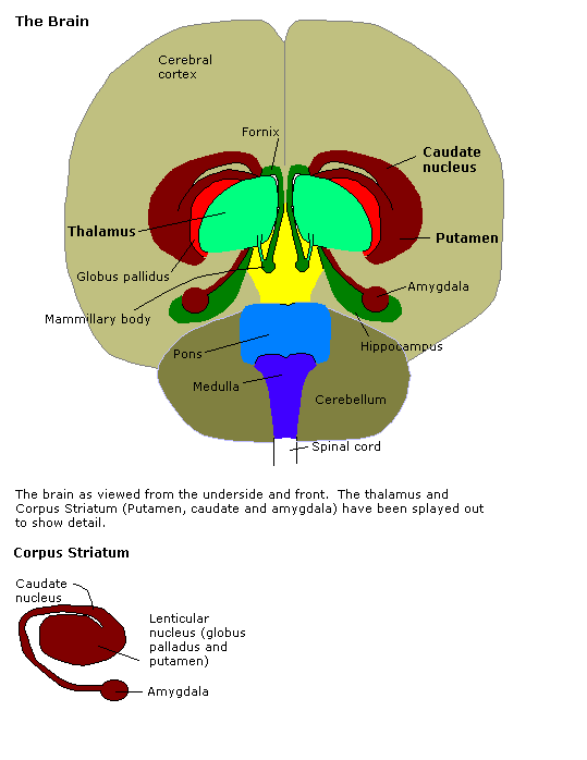
Size of the Caudate Nucleus
The caudate nucleus or caudatum is adjacent to the temporal lobes and looks like an elongated mass of grey matter. Because of its role in Parkinson’s disease, it has traditionally been associated with motor processes. It other roles include procedural learning, associative learning, inhibitory control of action, and it plays a role in the reward system.
Two studies on humans have shown the caudatum to be larger in females.
Size of the Cerebellum
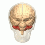
Found in the posterior portion of the brain in humans and other mammals, the cerebellum is distinguishable from the rest of the brain in colour and structure. It is important for controlling movement (coordination, precision, and accurate timing), and may also be involved in some cognitive functions such as attention and language, as well as regulating fear and pleasure responses. It integrates the data received from spinal cord and other parts of the brain to fine-tune motor activity. Damage to this part of the brain leads to disorders in fine movement, equilibrium, posture, motor learning, etc.
While studies show that the cerebellum is on average larger in males, once adjustment for overall brain size are made, the difference is eliminated.
Size and Composition of Hippocampus
The hippocampus, named so for its resemblance to the seahorse, is vital to spatial and cognitive memory functions.

In Alzheimer’s disease, this is one of the first regions of the brain to suffer damage, leading to short-term memory loss, disorientation as early symptoms.
![]()
Studies on humans conclude that it is larger in females — while studies on rodents indicate that it is larger in males. Size may be species-dependent as well as closely related to ecology.
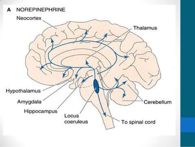
Size of Locus Coeruleus
The locus coeruleus is a small segment of the brain stem with a high concentration of neurons, and it is thought to play important roles in stress/anxiety and fear-response/panic. In the brain, it is the main site for the synthesis of norepinephrine.
(in the image below you can see the circulation of norepinephrine, also called noradrenaline, which mobilizes the brain and body for action under stress, danger, and fight-or-flight response. This substance increases arousal and alertness, promotes vigilance, enhances memory and focused attention, but also anxiety. It increase heart rate and blood pressure, triggers the release of glucose from energy stores, increases blood flow to skeletal muscles, reduces blood to the gastrointestinal system, and inhibits voiding the bladder… )
The only studies are on rodents indicate that is wider in females and longer in males, with no clear documentation of size difference.
Size of Olfactory Bulb
The olfactory bulb is important for interpreting odours. While no studies seem to exist in humans, in rats it is larger in males.

Size of Spinal Cord and Its Parts
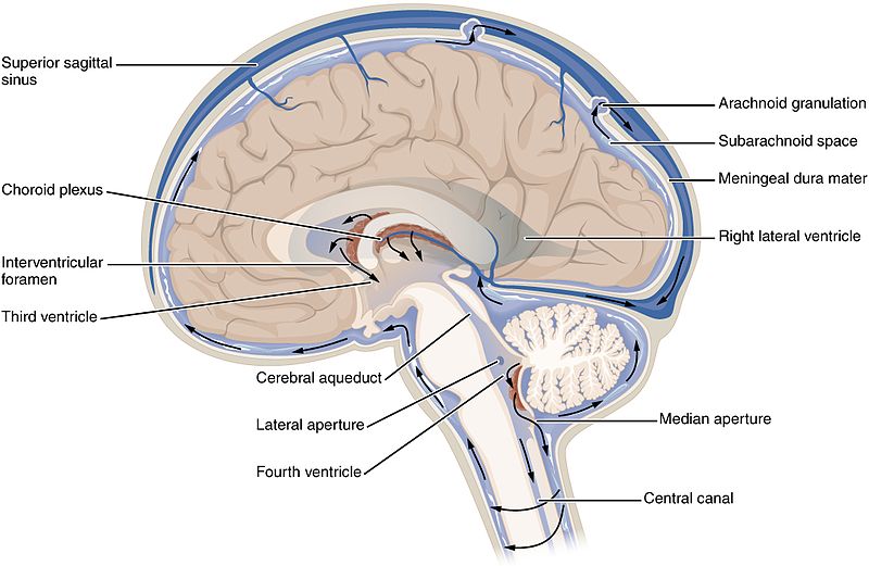
The spinal cord is the long, thin, tubular structure comprised of nervous tissue, extending from the medulla oblongata in the brainstem to the lumbar region of the vertebral column. The brain and the spinal cord together make up the central nervous system mentioned in the previous entry.

Within it is contained the central canal of the spinal cord that also contains the cerebrospinal fluid. The brain generates about 500 mL of this clear, colourless fluid every day, and it acts as a protection buffer to the brain both mechanically and immunologically, as well as playing other roles, including blood flow to the brain. (see the arrows in the image below to understand its circulation)
Studies show that the volume of the spinal nucleus innervating the bulbocavernous (or bulbospongiosus) muscle is greater in males.
Size of Striatum
The striatum, (or corpus striatum, neostriatum, striate nucleus), is a large division of the forebrain comprised of the caudate nucleus and the putamen, and which is rich with dopamine pathways. It plays a central role in movement, as well as the reward system. It coordinates multiple aspects of cognition, including motor and action planning, decision-making, motivation, reinforcement, and reward perception.

One study in Europe shows that it larger in males, while other studies show no difference between males and females.
Size of the Thalamus

The thalamus is a bundle of nerve cells of similar size and shape to an egg, located in the middle of the brain just above the brainstem. It has extensive nerve connections to both the midbrain and the cerebral cortex. It serves to process and route incoming motor and sensory stimuli to the various areas of the brain. It also regulates sleep, alertness and wakefulness.
Females have a larger thalamus than do males.
